Using the SuperFMX/Modality view
The modality or anatomical workflow (SuperFMX/Modality view), similar to what one experiences with DEXIS, is provided in Dentrix Ascend Imaging to help improve the functionality, user interface, and overall experience for users who are accustomed to using DEXIS and then switch to Dentrix Ascend Imaging. The SuperFMX/Modality view provides a level of flexibility and efficiency to imaging workflows. You can switch between the traditional template or exam-based workflow (Exam view) and the modality or anatomical workflow (SuperFMX/Modality View) as needed to accommodate your organization's diverse operational preferences and clinical needs and support your diagnostic processes.
Note: For information about switching to the SuperFMX/Modality view, refer to the topic about changing Dentrix Ascend Imaging settings.

As needed, you can filter images in the Modality view.
The tabs on the left navigation panel provide dedicated views for specific imaging modalities:
-
Intraoral X-ray - Displays all intraoral X-rays for the patient. This modality is selected by default. The FMX 18 anatomical view is displayed by default, with images organized by tooth number, and it provides a comprehensive overview of the patient's dental anatomy. There are options for switching to an FMX 21 anatomical view, using a tiled view, and flipping the view.
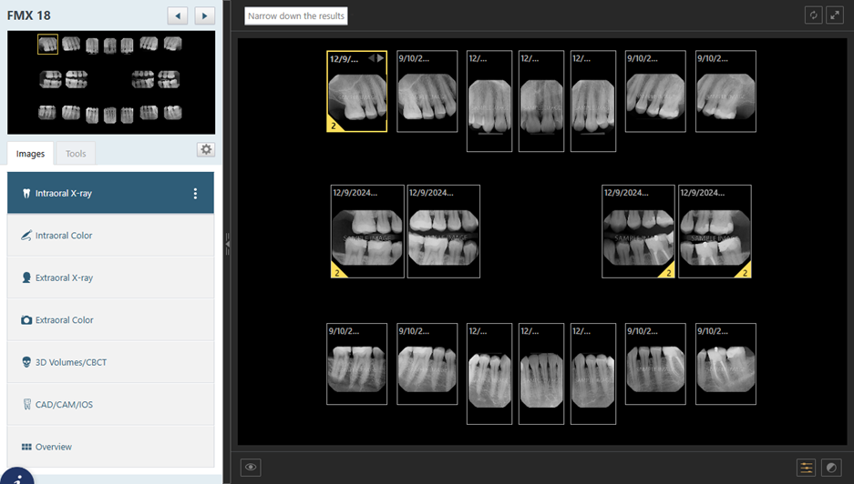
Notes:
-
Each image is assigned a slot based on the tooth number.
-
For a slot with multiple images, the newest image is displayed at the top of each stack. The yellow corner indicators on image stacks indicate that additional images are available.
-
For information about changing the view for this modality, refer to the topic about changing the view options for a modality.
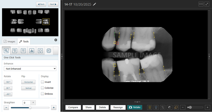
You can click (or tap) a selected image to view it in the single-image view, which provides an isolated, focused view of that image. The view includes a filmstrip navigator for scrolling through all images in the stack and enhanced navigation controls for quick image selection.
-
-
Intraoral Color - Displays all intraoral color images for the patient. The FMX 18 anatomical view is displayed by default, with images organized by tooth number. There are options for switching to an FMX 21 anatomical view, using a tiled view, and flipping the view.
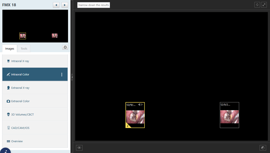
Notes:
-
Each image is assigned a slot based on the tooth number.
-
For a slot with multiple images, the newest image is displayed at the top of each stack. The yellow corner indicators on image stacks indicate that additional images are available.
-
For information about changing the view for this modality, refer to the topic about changing the view options for a modality.
You can click (or tap) a selected image to view it in the single-image view, which provides an isolated, focused view of that image. The view includes a filmstrip navigator for scrolling through all images in the stack and enhanced navigation controls for quick image selection.
-
-
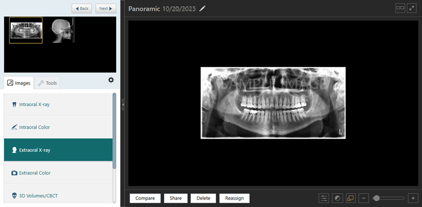
Extraoral X-ray - Displays all extraoral x-rays for the patient in a chronological, tiled view.
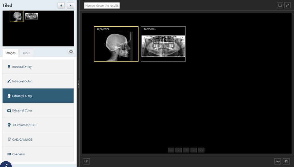
You can click (or tap) a selected image to view it in the single-image view, which provides access to a variety of tools for detailed analysis and editing, allowing for precise examination of extraoral radiographic data.
-
Extraoral Color - Display all extraoral color images for the patient in a chronological, tiled view.
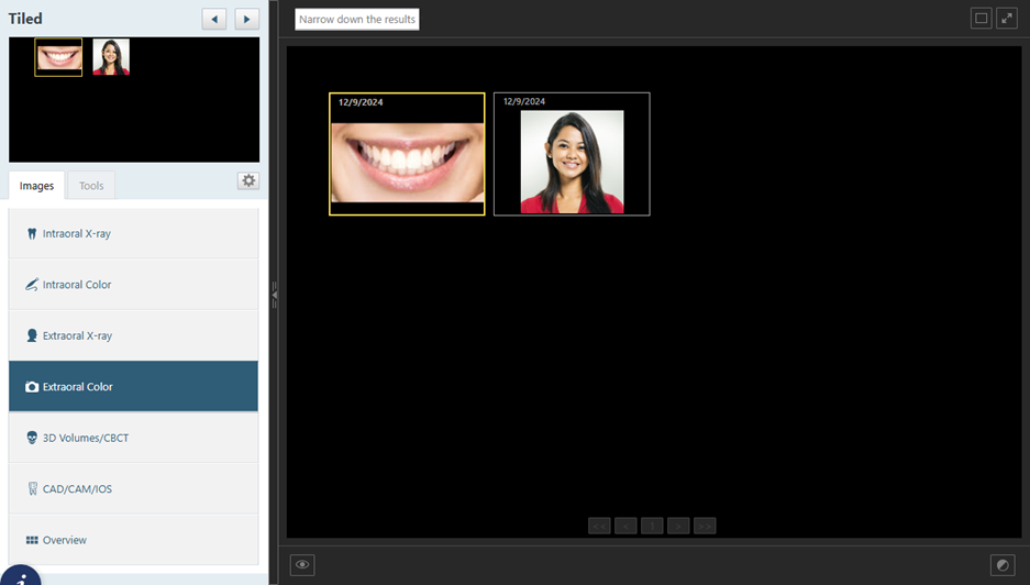
You can click (or tap) a selected image to view it in the single-image view, which access to a variety of tools for detailed analysis and editing, enhancing the examination of extraoral photographs.
-
3D Volumes/CBCT - Displays all 3D volumetric and cone beam computed tomography (CBCT) images for the patient in a chronological, tiled view.
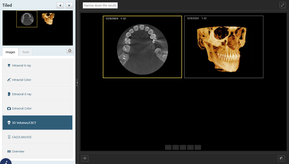
You can click (or tap) a selected image to view it in the single-image view, which provides access to a range of tools for detailed analysis and editing, allowing for precise examination of the volumetric data.
-
CAD/CAM/IOS - Displays all CAD/CAM and intraoral scanner (IOS) images for the patient in a chronological, tiled view.
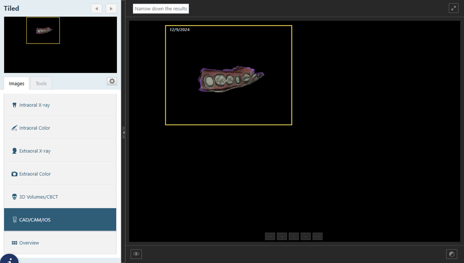
You can click (or tap) a selected image to view it in the single-image view, which provides access to a variety of tools for detailed analysis and editing.
-
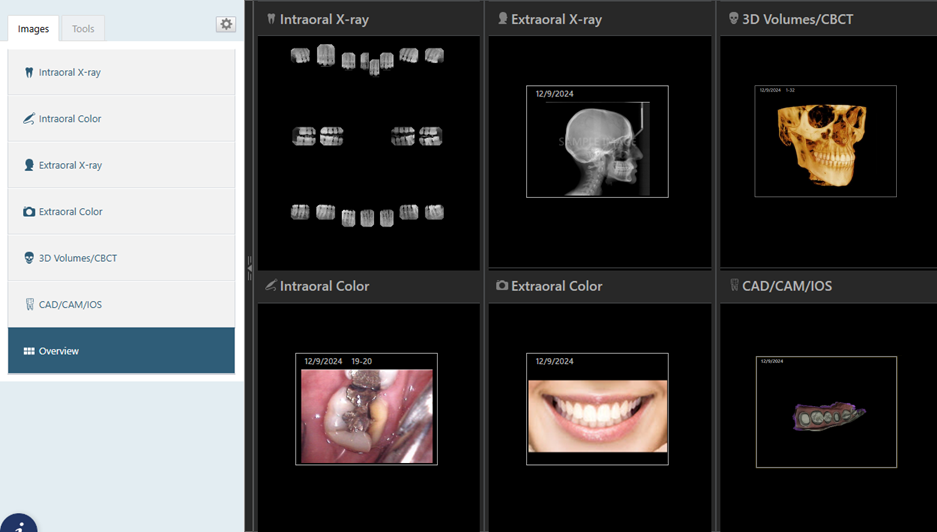
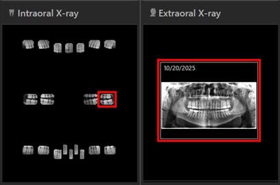
Overview - Shows a snapshot of all imaging modalities for the patient. Each modality is represented visually, allowing you to quickly assess available imaging data.
-
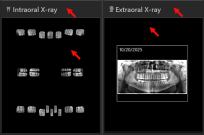
Clicking (or tapping) the heading of a modality's box or anywhere in the box where there is not an image redirects you to the corresponding modality tab, enabling seamless access to detailed views and further functionality.
-
Clicking (or tapping) one of the images in the Intraoral X-ray modality's box redirects you to the single-image view of that image with the Tools tab selected. Clicking (or tapping) an image in a different modality's box redirects you to the single-image view of that image on the corresponding modality tab.
-