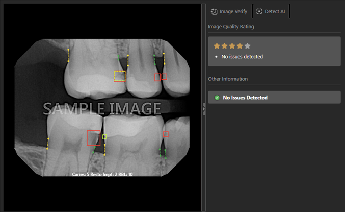
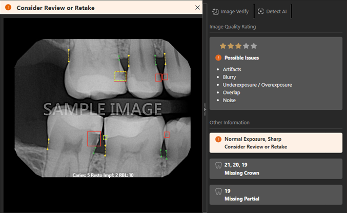
Viewing the image quality rating during acquisition
During the acquisition of a radiograph, Dentrix Ascend Imaging automatically assesses the quality of the image. The image is assigned a rating from one to five stars based on the overall diagnostic usability of the image. The rating reflects how suitable an image is for clinical interpretation and annotation. Although assessing image quality contains an inherent subjective component, you can rely on consistent visual criteria to ensure evaluation consistency.
Evaluations are based on the following criteria:
-
Sharpness and clarity
-
Contrast and exposure
-
Image noise
-
Motion blur
-
Anatomical coverage
-
Overlapping of anatomical structures (particularly contact points in bitewing radiographs)
-
Presence of artifacts
-
Visibility of key anatomical landmarks (CEJ, lamina dura, PDL space, apices)
The final rating reflects the overall diagnostic usability of the image, rather than the presence of a single isolated issue.
To view the image quality rating
Acquire an intraoral X-ray image.
One of the following occurs:
-
After you acquire an image in a series, the rating may appear at the top (according to the "Show quality star rating" setting). Also, the rating and a list of possible issues (if the rating is one to three stars) appear on the AI panel (on the right).
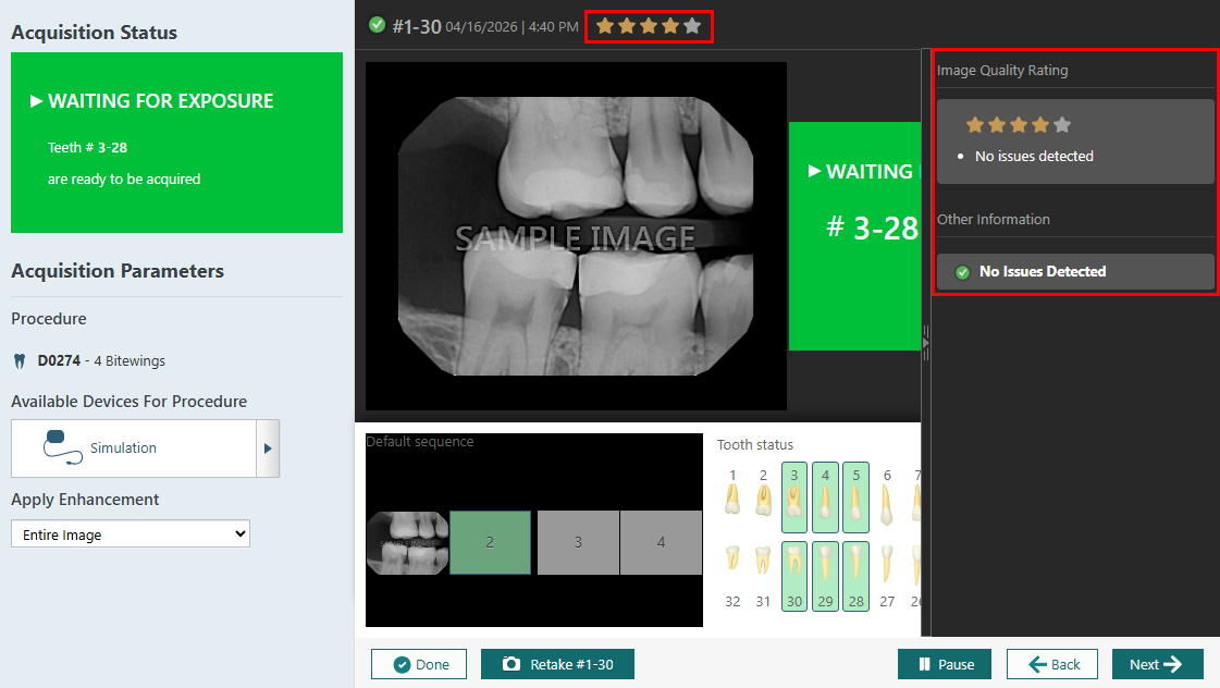
-
After you acquire an individual image, or after you acquire the last image in a series, when the image preview appears, the rating and a list of possible issues (if the rating is one to three stars) appear on the AI panel (on the right)
1 Star - Very poor quality
The image is non-diagnostic or almost non-diagnostic.
Characteristics:
-
Severe blur or motion artifact
-
Extreme under exposure or overexposure
-
Missing essential anatomical structures (for example, apices not visible in periapical radiographs)
-
Severe overlapping
-
Strong artifacts interfering with interpretation
The image cannot be reliably used for diagnostic assessment or annotation.
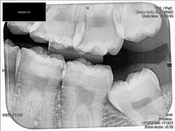
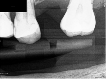
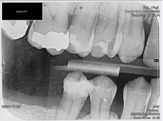
Examples of a 1-star rating
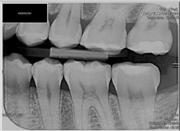
Severe overlapping
Severe underexposure
Motion blur present
2 stars - Poor quality
The image has significant limitations that reduce diagnostic confidence.
Characteristics:
-
Noticeable blur
-
Inadequate contrast
-
Partial anatomical cutoff
-
Marked overlapping
-
Moderate artifacts
Interpretation is possible but limited and associated with lower confidence.
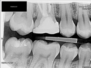
Examples of a 2-star rating
Marked overlapping
Noticeable blur
3 Stars - Acceptable quality
The image is diagnostically usable but not optimal.
Characteristics:
-
Minor blur
-
Slight exposure imbalance
-
Limited overlapping
-
Mild noise
-
Small artifacts that do not significantly interfere
All essential anatomical structures are visible. Annotation and pathology detection can be performed with reasonable confidence.
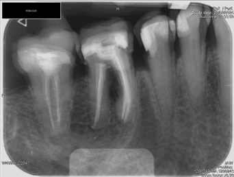
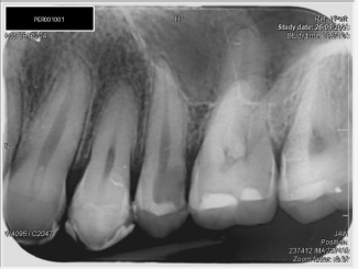
Examples of a 3-star rating
Small artifacts, slightly underexposed, limited overlapping
Slightly underexposed, limited overlapping
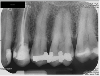
4 Stars - Good quality
The image shows good clarity and is well-suited for interpretation.
Characteristics:
-
Good sharpness
-
Balanced exposure and contrast
-
Minimal overlapping
-
No significant artifacts
-
Clear visualization of relevant anatomical structures
Minor imperfections may be present but do not affect diagnostic performance.
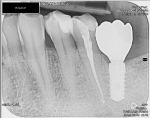
Examples of a 4-star rating
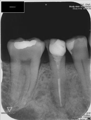
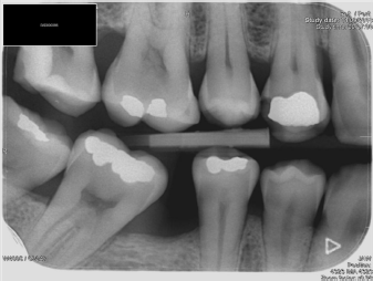
5 Stars - Excellent quality
The image demonstrates optimal diagnostic quality.
Characteristics:
-
Excellent sharpness and resolution
-
Proper exposure and contrast
-
No relevant artifacts
-
Complete anatomical coverage
-
Clear visualization of fine details (for example, lamina dura, PDL space)
Image allows high-confidence interpretation and precise annotation.
Examples of a 5-star rating