Using the AI panel

When you view an image in Dentrix Ascend Imaging, an AI panel appears. The panel displays the following information:

  • On the Magnify tab, zoomed-in views of AI findings (for caries and PRL only) appear.

  • On the Image Verify tab, a star-based image quality rating appears with contextual issue messaging.

  • On the Detect AI tab, a summary of the AI analysis performed on the displayed teeth appears. This tab is available only if Detect AI has been activated, and the image being viewed is eligible for AI findings.

Note: Depending on the width of the AI panel, the tabs names may be hidden, leaving only icons to identify the tabs.

To use the AI panel

  1. If the correct patient is not already selected, use the Patient Search box to access the patient's record.

  2. Do one of the following:

    • View an image from the patient's image history list (Exam view).

      1. On the Patient menu, under Clinical, select Chart, Progress Notes, Quick Exam, Perio, Tx Planner, or Imaging.

        The patient's clinical record opens with the Chart, Progress Notes, Quick Exam, Perio, Tx Planner, or Imaging tab selected.

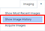
      2. On the Imaging tab's menu, select Show Image History.
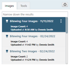
      3. On the Images tab, select an exam for an individual image or a series.
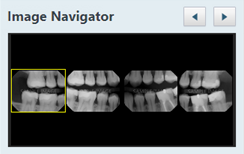
      4. If you have selected a series, under Image Navigator, click (or tap) an image of that series.
    • View an image from the patient's image history list (Modality view).

      1. On the Patient menu, under Clinical, select Chart, Progress Notes, Quick Exam, Perio, Tx Planner, or Imaging.

        The patient's clinical record opens with the Chart, Progress Notes, Quick Exam, Perio, Tx Planner, or Imaging tab selected.

      2. On the Imaging tab's menu, select Show Image History.

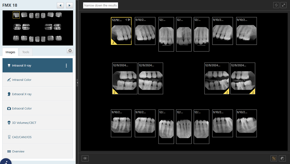
      3. On the Intraoral X-ray tab, select a slot of the template, and then click (or tap) it again.
    • View one of the patient's most recent images.

      1. Do one of the following:

        • On the Patient menu, under Clinical, select Imaging.

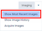
        • On the Patient menu, under Clinical, select Chart, Progress Notes, Quick Exam, Perio, Tx Planner, or Imaging. Then, on the Imaging tab's menu, select Show Most Recent Images.
      2. Click (or tap) an individual image or an image in a series.
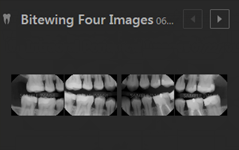
    The selected image appears in the viewing area.

    Notes:

    • The AI panel is visible or hidden by default according to the "Show right panel on launch" setting.

    • If the panel is visible, to hide the AI panel, click (or tap) the bar on the left edge of the panel.

    • If the panel is hidden, to show the AI panel, click (or tap) the bar on the left edge of the panel.

    • If you have collapsed or expanded the AI panel, the panel remains collapsed or expanded as you switch between a patient's exams.

    • Whichever tab you have selected on the AI panel remains selected as you switch between a patient's exams.

  3. View information and perform actions as needed on any of the following tabs of the AI panel:

    • Magnify - On this tab, zoomed-in views of AI findings (for caries and PRL only) appear. This tab displays a magnified view for each AI finding on the image being viewed, allowing you to quickly assess areas of concern without having to manually zoom in.

      • If you position your pointer over an AI finding on an image in the viewing area, the corresponding magnified view on the Magnify tab is selected automatically (a yellow indicator box appears around that magnified view).

      • If you position your pointer over a magnified view on the Magnify tab, the corresponding AI finding on the image in the viewing area is selected automatically (a yellow indicator box appears around that AI finding).

      Notes:

      • The Magnify tab is available only if Detect AI is enabled and you are viewing an intraoral X-ray image that is eligible for AI findings.

      • If any tool besides "One Click Tools" is active, the Magnify tab does not display magnified views.

      • If you apply an enhancement (Enhance, Invert, Colorize, or Emboss) to an image in the viewer, the same enhancement is applied to the images on the Magnify tab.

      • If you adjust the brightness or contrast of an image in the viewer, the same adjustment is applied to the images on the Magnify tab.

      • If AI findings are turned off, the Magnify tab does not display magnified views.

    • Image Verify - On this tab, a star-based image quality rating appears with contextual issue messaging so you can quickly and confidently assess image quality without ambiguous guidance. Based on the star rating of an image, the messaging that appears clearly explains the detected quality concerns or confirms that no issues were found.

      • An image with possible issues detected:

        Notes:

        • If the Image Verify tab is selected on the AI panel, while you are viewing an image that has a 3-star rating or less, the "Image Quality Insight Available" banner may appear above the image (according to the "Show quality star rating" setting). This holds true whether the AI panel is currently visible or hidden.

        • If you dismiss the "Image Quality Insight Available" banner, it remains dismissed as you switch between a patient's exams.

      • An image with no issues detected:

      The following Image Quality Rating may appear:

      • A star-based rating from 1 to 5 stars.

      • Depending on the rating, the applicable messaging appears:

        • For a 1-, 2-, or 3-star rating, the following list of Possible Issues appears:

          • Artifacts

          • Blurry

          • Underexposure / Overexposure

          • Noise

          • Contrast

        • For a 4- or 5-star rating, the following text appears:

          • No Issues Detected

    • Detect AI - On this tab, a summary of the AI analysis performed on the displayed teeth (using all images, with those same teeth in common, that were acquired during that visit—in the same series) appears.

      • The odontogram at the top displays the teeth that may require restorative procedures in a purple color. Position your pointer over any tooth to see the tooth number and "Restorative" (if applicable).

      • The AI Summary displays damage levels per tooth and bone levels per quadrant:

        • For Tooth Damage, if not all damaged teeth appear, to view the entire list, click (or tap) the Show all link.

        • For Bone Level, to view distal and mesial bone level measurements for each each tooth in each quadrant, click (or tap) the Show measurements link.

      Do any of the following as needed:

      • View patient education topics to provide visual aids that may help the patient understand his or her treatment needs:

        1. Click (or tap) Patient Education.

          The Patient Education dialog box appears.

        2. Select Adult or Pediatric, depending on the patient's age and dentition.

        3. Select a treatment:

          • Visual aids for the following adult treatments are available:

            • Filling

            • Root Canal

            • SRP

            • Crown

            • Tooth Enamel Repair

            • Implant

          • Visual aids for the following pediatric treatments are available:

            • Fluoride (Preventative)

            • Tooth Enamel Repair (Incipient Lesion)

            • Stainless Steel/Zirconia Crown

            • Space Maintainer

            • Sealant

      • Share the AI summary and patient education topics with the patient:

        • To provide a QR code, do the following:

          1. Click (or tap) Share with Patient, and then select QR Code.

            The Patient Education Summary dialog box appears.

          2. Click (or tap) Edit More to customize the report and generate a new QR code.

            1. Set up the following options:

              • From the Select Teeth list, select the checkbox of each tooth to include.

              • Under Configurations, set up the following options:

                • Treatments - Select the checkbox of each patient education topic to include.

                • Note - Enter notes to include.

                • AI Findings to Include - Select the checkbox of each type of finding to include.

            2. Click (or tap) Generate QR Code.

          3. Have the patient scan the QR code.

        • To provide a downloaded file, do the following:

          1. Click (or tap) Share with Patient, and then select Print/Download.

            The Patient Education Summary dialog box appears.

          2. Customize the report:

            • From the Select Teeth list, select the checkbox of each tooth to include.

            • Under Configurations, set up the following options:

              • Treatments - Select the checkbox of each patient education topic to include.

              • Note - Enter notes to include.

              • AI Findings to Include - Select the checkbox of each type of finding to include.

          3. Click (or tap) Download. The report is saved as a .jpg file.

          4. Send the report to the patient.

        • To provide a printed document, do the following:

          1. Click (or tap) Share with Patient, and then select Print/Download.

            The Patient Education Summary dialog box appears.

          2. Customize the report:

            • From the Select Teeth list, select the checkbox of each tooth to include.

            • Under Configurations, set up the following options:

              • Treatments - Select the checkbox of each patient education topic to include.

              • Note - Enter notes to include.

              • AI Findings to Include - Select the checkbox of each type of finding to include.

          3. Click (or tap) Print.

          4. Use the browser's Print dialog box to print the report.

          5. Give the printout to the patient.