

Henry Schein One has partnered with VideaHealth to apply artificial intelligence (AI) analysis to new and existing bitewings and periapical X-rays in Smart Image. Dentrix Detect AI helps you with the following FDA approved features:
Detecting caries in the enamel (dotted line yellow rectangles) and dentin (solid line red rectangles) on all primary and secondary teeth of patients three years and older. This eliminates the need for manual analysis of all primary and secondary teeth, saving you and your hygienists time.
Using Dentrix Detect AI to analyze periapical X-rays can assist you in detecting caries on anterior teeth. This can lead to earlier intervention and better patient outcomes.
Measuring interproximal Radiographic Bone Levels in bitewings and Periapical Radiographs (PAs) for patients who are at least 12 years old. Bone level detection is only possible with mesial and distal surfaces. RBL measurements appear as green, yellow, orange, or red dotted lines.
Detecting periapical radiolucency (PRL) for patients 22 years and older to more effectively identify bone demineralization due to infections around root apices, cysts, and other causes. PRL is indicated by a red circle or oval.
Detecting interproximal calculus to educate your patients 12 years and older on the importance of scaling and root planing. Combined with measuring interproximal Radiographic Bone Levels, Dentrix Detect AI illustrates disease prognosis for the patient and clinician. Interproximal calculus is indicated by an orange circle or oval.
Detecting restoration imperfections in patients 22 years and older by identifying imperfect crown and filling margins and voids. A restoration imperfection is indicated by a yellow rectangle.
To view additional Dentrix Detect AI indications
1. In the Smart Image toolbar, click the Acquire a 2D/3D or CAD/CAM Image button.
The Smart Image Acquisition dialog box appears.
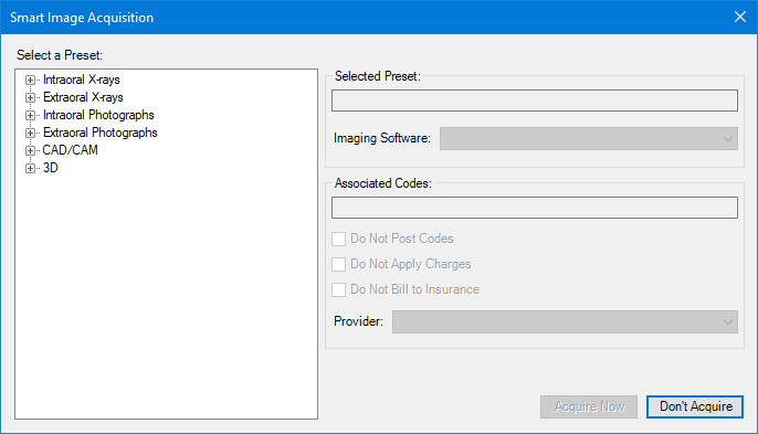
2. Under Select a Preset, click the image type (Intraoral X-rays), and then the specific type of image (1BW) you want to capture.
The type of image you selected appears in the Selected Preset text box, and if applicable, the associated codes in the Associated Codes text box.
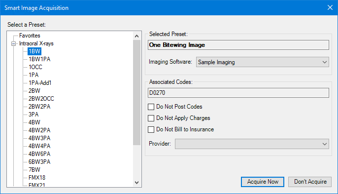
3. From the Imaging Software list, select the imaging software that you want to open.
4. Select one of the following options:
Do Not Post Codes – No procedure codes are posted for the acquisition.
Do Not Apply Charges – The amount of posted procedure codes is set to $0.
Do Not Bill to Insurance – Posted procedure codes are not billed to insurance providers.
5. To change the provider, click the Down arrow, and select the appropriate provider from the list.
6. Click Acquire Now.
The imaging application opens from which you can select the template and acquire the images. The images are automatically submitted to VideaHealth. The Dentrix Detect AI analyzed images appear in the Smart Image panel.
In Figure 1, caries are indicated by red squares, RBL is indicated by vertical dotted lines, calculus is indicated by an orange circle, and restorative imperfections are indicated by yellow rectangles.
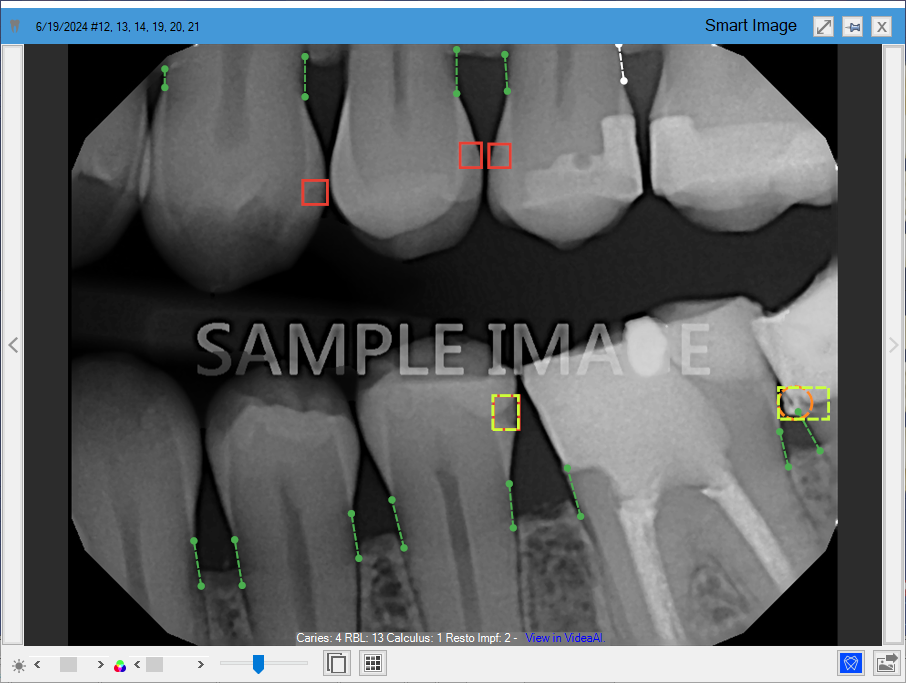
Figure 1. Diagnostic Viewer with Dentrix Detect AI indications.
In Figure 2, caries are indicated by red rectangles, RBL is indicated by vertical dotted lines, and restorative imperfections are indicated by yellow rectangles and squares.
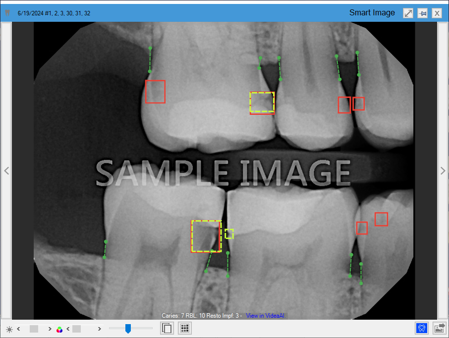
Figure 2. Diagnostic Viewer with Dentrix Detect AI indications.
In Figure 3, caries are indicated by a red square, RBL is indicated by vertical dotted lines, PRL is indicated by red ovals, and restorative imperfections are indicated by yellow rectangles.
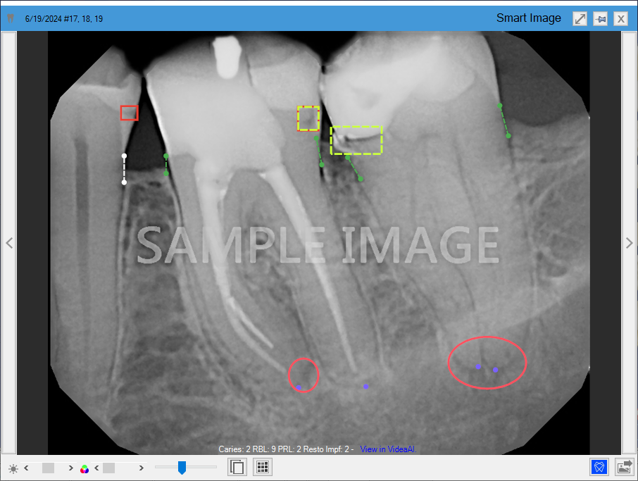
Figure 3. Diagnostic Viewer with Dentrix Detect AI indications.